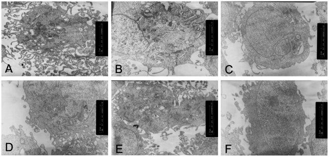
Figure 3. Transmission electron microscopic analysis of the HK-2 cells cultured under different conditions.
(A–C)The HK-2 cells under 30 mM glucose (B) or 30 mM glucose+Cont siRNA (C) showed decreased number of microvilli, mitochondria and increased volume density of rough endoplasmic reticulum compared to that of the cells cultured in 5.5 mM glucose(A). (D–F)Changes of the cells were reversed,which were cultured in the 30 mM glucose+p38 siRNA for 24 h(D) or 48 h(E) or 30 mM glucose+AP-1 inhibitor(F).

