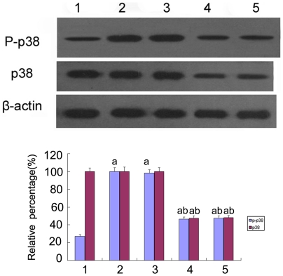
Figure 7. The expression of p38 MAPK and phosphorylated P38 MAPK by Western blot analysis.
p38 MAPK and phosphorylated p38 MAPK were measured by Western blot of the HK-2 cells cultured in 5.5 mM glucose (lane 1), 30 mM glucose (lane 2), 30 mM glucose+Cont siRNA(lane 3), 30 mM glucose+p38 siRNA for 24 h(lane 4), 30 mM glucose+p38 siRNA for 48 h(lane 5). Values represent the mean ± SD, aP<0.05 vs. 5.5 mM glucose, bP<0.05 vs. 30 mM glucose.

