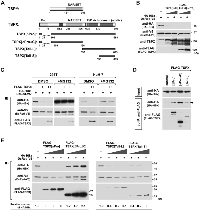
Figure 1. TPSX stimulates degradation of HBx in a proteasome dependent manner.
(A) Structure of TSPY, TSPX, and schematic representation of the truncated mutants of TSPX used in present study. (B) Effect of co-expression with TSPX on HBx stability in mammalian cells. 293T cells were co-transfected with HA-HBx expression vector (0.2 µg/well) in the presence or absence of TSPX[full] or FLAG-TSPX[ΔPro] expression vector (0.025, 0.1 µg/well). DsRed-V5 expression vector (0.1 µg/well) was co-transfected as the internal control for monitoring the transfection efficiency. Forty-eight hours after transfection, cells were lysed and analyzed by Western-blot (immuno-blot = IB) using indicated antibodies. (C) Effect of a proteasome inhibitor MG132 on TSPX-enhanced HBx degradation. 293T cells and HuH-7 cells were co-transfected with HA-HBx expression vector (0.2 µg/well) in the presence or absence of FLAG-TSPX[ΔPro] expression vector (0.025, 0.1 µg/well). DsRed-V5 expression vector (0.1 µg/well) was co-transfected similarly as above. Twenty-four hours after transfection, cells were treated with vehicle (DMSO) or 25 µM MG132 for additional 24 h. Cells were lysed and analyzed by Western-blot using indicated antibodies. (D) Interaction of TSPX and HBx in mammalian cells. HA-HBx expression vector was co-transfected into 293T cells with expression vector of FLAG-epitope tagged TSPX mutants. Twenty-four hours after transfection, cells were treated with 20 µM MG132 for additional 24 h. Coimmunoprecipitation was performed with anti-FLAG antibody, and immunoprecipitated complexes (co-IP) were analyzed by Western blot using anti-HA and anti-FLAG antibodies. One percent of each lysate (input) was analyzed in parallel as a transfection control. (E) Mapping of the functional domain for stimulation of HBx-degradation. HA-HBx expression vector (0.1 µg/well) was co-transfected into 293T cells with FLAG-TSPX[ΔPro] (0.05, 0.1, 0.2 µg/well), FLAG-TSPX[ΔProΔC] (0.05, 0.1, 0.2 µg/well), FLAG-TSPX[Tail-L] (0.05, 0.1, 0.2 µg/well) or FLAG-TSPX[Tail-S] (0.05, 0.1, 0.2 µg/well), and analyzed as described above. The results indicate that the D/E-rich C-terminal region is sufficient to promote HBx degradation.

