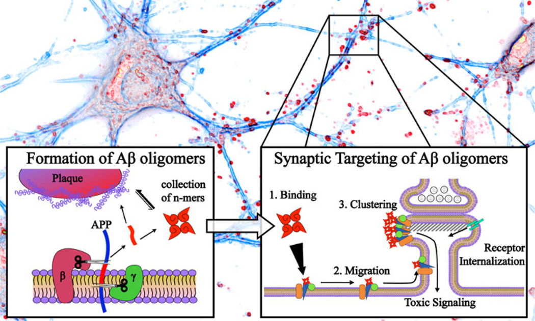
Fig. 1.
Formation and synaptic targeting of Aβ oligomers. A neuron is represented with blue neurites and red dendritic spines (background image). Following APP cleavage at the neural membrane by β- and γ-secretase, the 42-residue Aβ peptide (red) can be deposited into senile plaques or can alternatively form a collection of oligomeric species (left inset). Some component of this collection of oligomers interacts with neurons to form synaptic clusters which are associated with a variety of synaptic pathologies including the internalization of receptors involved in synapse function and maintenance and the initiation of toxic signaling cascades (right inset)

