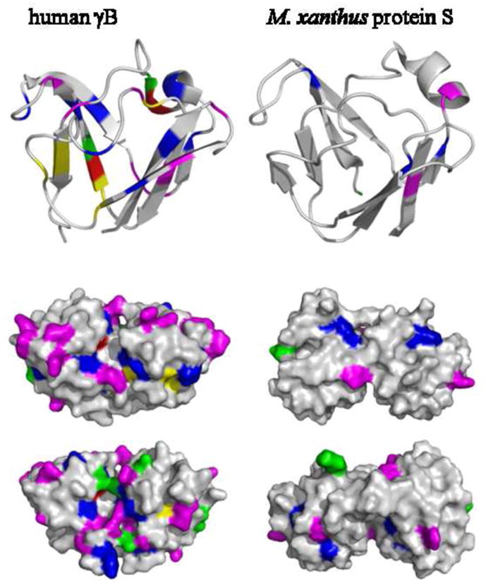
Figure 5.

Top: Ribbon representation of the N-terminal domain of human γB-crystallin (2JDF) and M. xanthus protein S (1PRR). Highlighted residues are in high relative abundance in γB-crystallin with high refractive index increment: methionine (green), arginine (magenta), cysteine (yellow), tryptophan (red), and tyrosine (blue). Middle and Bottom: Surface representation of the whole molecules containing two domains, viewed from two opposite angles.
