
Figure 6.
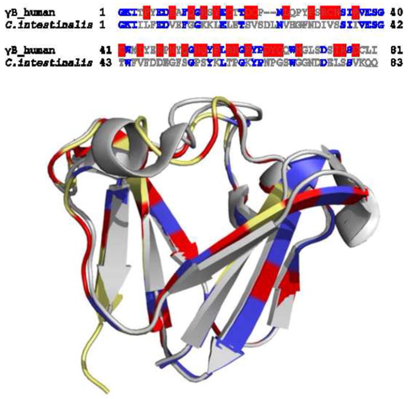
Sequence and structure comparison of N-terminal domain of human γB crystallin (2JDF) and βγ-crystallin from C. intestinalis (2BV2). (A) Sequence alignment of the two polypeptides. The identical residues are in blue, and the residues with higher dn/dc in human γB crystallin are highlighted in red. (B) Structural overlay of the two constructs. The N-terminal domain of human γB crystalline is in yellow and βγ-crystallin from C. intestinalis is in gray. The residues highlighted in blue are the identical ones. The residues in human γB crystalline which have higher dn/dc values compared to those at the same positions in βγ-crystallin C. intestinalis are highlighted in red.