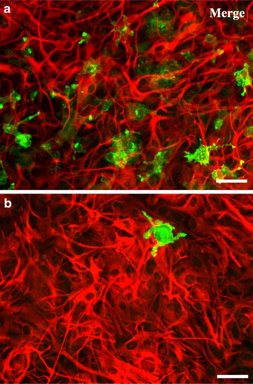
Fig. 1.
Characterization of primary astroglial cultures containing different percentage of microglia. Astrocytes were labelled with anti-GFAP (TRITC, red) and microglia with anti-CD11b (Alexa Fluor 488, green). Representative immunofluorescent micrographs of the two types of cultures: a co-cultures and b highly enriched astroglial cultures, double-labelled for GFAP and CD11b. In co-cultures, the number of microglia present was 8.0 ± 0.8% (n = 5) and in highly enriched astroglial cultures was 1.0 ± 0.3% (n = 5). Scale bar: 50 μm

