
Figure 4.
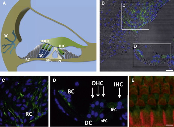
Localization of Smpx in the Mouse Inner Ear
(A) A schematic representation of the cochlea. The sensory epithelium is composed of inner hair cells (IHC) and outer hair cells (OHC). Nonsensory supporting cells include Deiters cells (DC), Böttcher cells (BC), and inner and outer pillar cells (iPC, oPC). Root cells (RC) build up a cellular network in the lateral wall of the cochlea.
(B) Cryosections (7-μm thickness) from the cochlea of adult C57BL/6J mice were stained with an Smpx antibody (Sigma Aldrich #AV41597, 1:500, green), and DAPI (blue). The overlay with differential interference contrast is shown. The scale bar represents 25μm. Stainings of SMPX_myc overexpressing HeLa cells with the Smpx rabbit-antiserum gave the same signal as with the mouse Myc-antibody (data not shown).
(C and D) Enlargements of spiral ligament cells and outer sulcus cells together with root cells (C) and the sensory epithelium with supporting cells (D). Note the Smpx localization in root cells, Böttcher cells, inner and outer pillar cell, and weaker signals in Deiters cells and hair cells (marked by arrows).
(E) Apical basilar membranes of fixed cochleae were dissected and immunostained with an Smpx antibody (green) and conjugated phalloidin (red). Weak Smpx immunoreactivity was detected in inner and outer hair cells. The scale bar indicates 5μm.