
Figure 1.
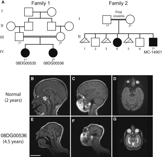
Pedigrees and Radiographic Features of the Two Consanguineous Families with Microlissencephaly
(A) Both families are from Saudi Arabia. Parents of family 1 are first cousins and have two affected female children (08DG00535, IV-1 and 08DG00536, IV-2). Whole-blood DNA from both parents and both affected children was obtained and analyzed (indicated by an asterisk). Parents of family 2 are first cousins who had seven reported pregnancies, producing one affected male (MC-14901, II-7), one affected female (II-4) (not available for analysis), two unaffected males, and three pregnancies that resulted in fetal demise. Whole-blood DNA from both parents and the affected male (MC-14901, II-7) was obtained and analyzed (indicated by an asterisk).
(B–G) Representative MRI images of 08DG00536 (IV-2) from family 1 at 4.5 years of age, demonstrating the drastic reduction in brain size (E–G), agenesis of the corpus callosum, and abnormal gyral pattern compared to that seen in a normal 2-year-old child (B-D). Sagittal T1 (B, C, E, and F) and axial T2 (D and G) sections are shown. The scale bar represents 5 cm. Additional images are available as Movies S1–S6.