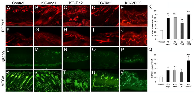
Figure 3. Cutaneous nerves are increased in number in KC-Ang1, KC-Tie2, EC-Tie2, and KC-VEGF skin compared to CD1 control animals and pattern similarly to cutaneous vessels.
Cutaneous nerves stained with the pan peripheral nerve marker PGP9.5 in (A, F) control, (B, G) KC-Ang1, (C, H) KC-Tie2, (D, I) EC-Tie2, and (E, J) KC-VEGF mice. Low magnification (A-E) images of thick skin (50μm) demonstrate PGP9.5+ nerve patterning throughout the dermis and epidermis and high magnification (F–J) images at the dermal-epidermal junction illustrate epidermal-specific nerve staining. Quantification of the number of nerves in each mouse strain (K) reveals all transgenic mice have more PGP9.5+ cutaneous nerves than control mice and KC-Tie2 and KC-VEGF animals have significantly more nerves than EC-Tie2 animals. Thin (8um) skin sections stained with antibodies targeting neurofilament 200 (NF200), a non-C fiber sensory nerves marker and quantitation demonstrates increases in NF200+ nerve fibers in all transgenic mice compared to control animals (L–Q). KC-VEGF mice have significantly more NF200+ nerves than KC-Ang1, KC-Tie2 and EC-Tie2 mice. Back skin sections stained with MECA antibodies (R–V) demonstrate moderate similarities between vascular and neural patterning as compared with the PGP9.5+ stained sections. * p< 0.05 compared to control animals; + p<0.05 compared to EC-Tie2 mice; ** p<0.05 compared to KC-Ang1, KC-Tie2 and EC-Tie2 mice.

