Figure 5.
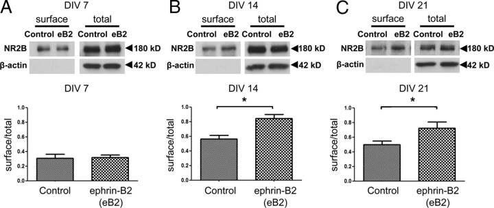
Ephrin-B2 activation of EphB2 increases NR2B surface localization. A–C, Cortical neurons at 7 DIV (A), 14 DIV (B), or 21 DIV (C) were treated for 45 min with control Fc (C) or activated ephrin-B2-Fc (eB2). Biotinylated (surface) and total NR2B protein was visualized by immunoblotting with specific antibodies (top gels). β-Actin was used as a loading control for total protein (bottom gels). Absence of actin in surface (biotinylated) gels indicates validity of surface labeling. Representative immunoblots show no actin immunolabeling in the biotinylated surface fraction. The bottom bar graphs show the ratio of amount of surface NR2B to total NR2B at 7 DIV (n = 5 experiments), 14 DIV (n = 6 experiments), or 21 DIV (n = 6 experiments). Ephrin-B2-Fc versus Fc (control) conditions were analyzed by an unpaired t test. *p < 0.05. Error bars indicate SEM.

