
Fig. 4.
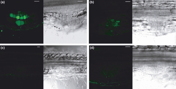
Detection of nitric oxide (NO) in nodule primordium by confocal laser scanning microscopy. DAF-2DA fluorescence (green) was observed on fresh slices (100 μm) of the root infection zone. The roots slices on (b) and (d) were pretreated with 1 mM of cPTIO (2-(4-carboxyphenyl)-4,4,5,5-tetramethyl imidazoline-1-oxyl-3-oxide) for 45 min before the treatment with the DAF-2DA probe. Each picture is composed by the DAF fluorescence and transmission micrographs. In pictures (a) and (b), DAF fluorescence was observed in dividing cells of the nodule primordium. The cPTIO treatment reduced significantly the fluorescence (c,d) which confirmed the specificity of the fluorescence. Root slices analysed without incubation with the DAF-2DA probe did not show autofluorescence in our acquisition settings. The typical fluorescence emission spectrum of the DAF probe was also confirmed (Data not shown). Data are from four independent experiments (n=48) and pictures shown are representative of the different conditions. Bars, 50 μm.