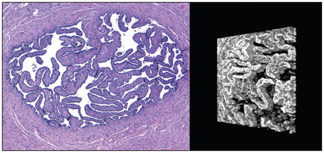
Figure 1.
Side-by-side views of normal fallopian tube lumen. (Left) A hematoxylin–eosin stained section at 4× objective magnification. The tube wall is composed of smooth muscle tissue. The lumen contains primary folds lined by epithelial cells. (Right) A three-dimensional reconstruction of a spectral domain optical coherence tomography scan of the tissue block, turned slightly (lumen only). Primary folds are visible as three-dimensional structures.

