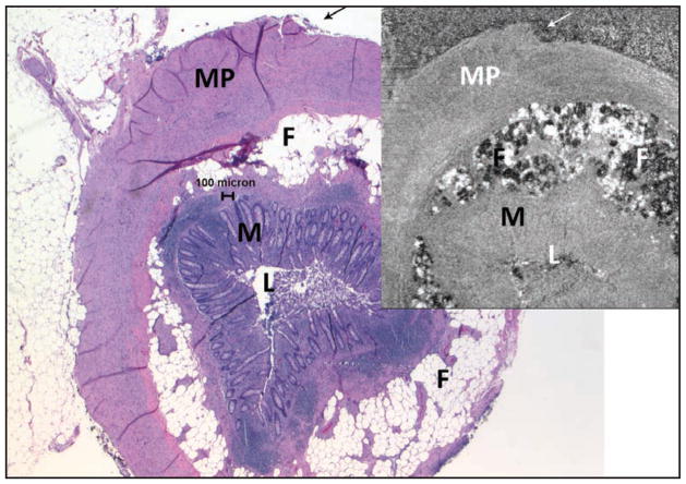
Figure 7.
Normal appendix (larger image, hematoxylin–eosin at 2× objective magnification, upper-right spectral domain optical coherence tomography [SD-OCT] 3 × 3 mm). These images were matched using the outline of the tissue (arrows) and other tissue patterns. Fat (F) lobules are both light and dark; muscularis propria (MP), mucosa (M), and lumen (L) are also evident. Note that the SD-OCT image in this figure was flipped horizontally to match the histologic section. It is uncommon but not rare for a histotechnologist to inadvertently “flip” a tissue section so that the slide is a mirror image of the block, or even of other slides from the same block.

