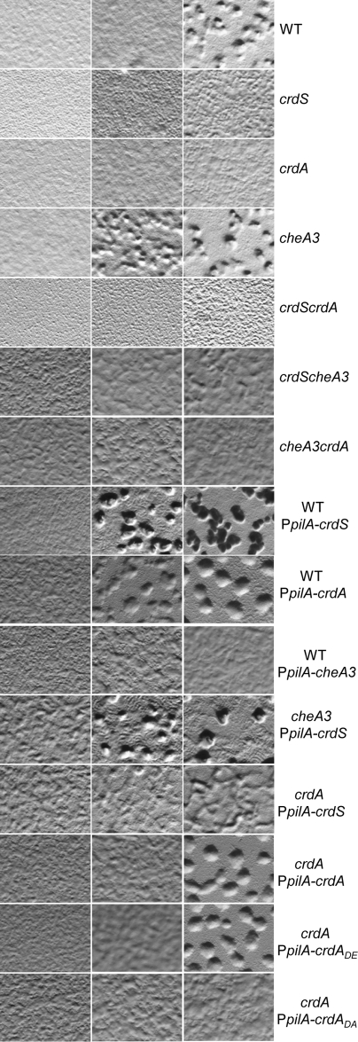
FIG 2 .
crdS, cheA3, and crdA mutants display altered timings of development. Developmental assays were performed as described in Materials and Methods. Phenotypic assays were conducted by spotting 10 µl of cells at 250 Klett units on starvation (CF) media. Images were acquired at 50× magnification at 18, 24, and 48 hours (left to right). Developmental progression is indicated by the presence of aggregates or opaque fruiting bodies containing 105 to 106 cells. The constitutively active pilA promoter (PpilA) was used to express crdS from the ectopic Mx8 phage attachment site. Premature or delayed phenotypes are relative to the wild-type parent.

