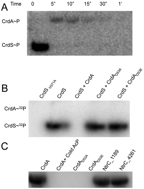
FIG 4 .
CrdS phosphorylation of CrdA in vitro. (A) Phosphotransfer between CrdSsoluble~32P and CrdA. Loss of phosphorylated CrdS indicates rapid phosphotransfer to CrdA. Complete transfer occurs within 5 seconds, as indicated by the loss of the CrdSsoluble~P band and the appearance of the CrdA~P band. (B) CrdS(H371) and CrdA(D53) are required for phosphotransfer. All reaction mixtures contain excess total ATP and 0.3 µM [γ-32P]ATP in kinase buffer and were incubated for 30 minutes, fractionated by SDS-PAGE, and visualized by autoradiography as described. Lane 1 contains CrdS(H371A), which is unable to autophosphorylate. Lane 2 contains phosphorylated CrdSsoluble. Lane 3 contains CrdSsoluble incubated with CrdA for 10 minutes, leading to complete loss of CrdS. CrdSsoluble~P is unable to transfer phosphoryl groups to CrdA(D53A) (lane 4) or CrdA(D53E) (lane 5). (C) CrdA is phosphorylated by acetyl phosphate (AcP). Radiolabeled AcP was generated as described in Materials and Methods. When CrdA is incubated with [32P]AcP, CrdA~32P is formed (lane 1). When the conserved residue, D53, of CrdA is mutated [to CrdA(D53A) or CrdA(D53E)] or when wild-type CrdA is incubated with excess unlabeled AcP (Sigma), no labeling is apparent (lanes 2 to 4). The alternative target proteins, NtrC_1189 and NtrC_4261, were phosphorylated with radiolabeled AcP (lanes 5 and 6).

