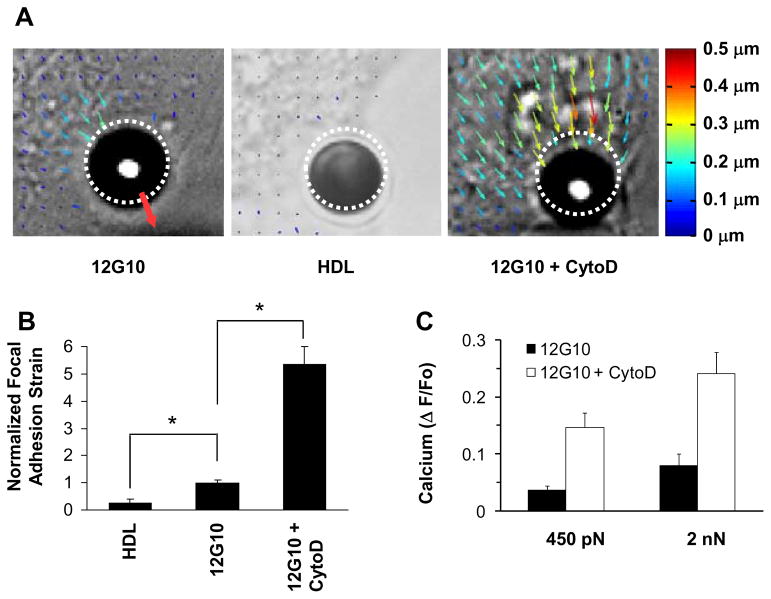
Fig. 2. Activation of calcium signaling by mechanical strain within focal adhesions.
(A) Maps of cytoplasmic displacement directly beneath bound microbeads immediately after application of a force pulse (2000 pN, 500 msec). Dashed circles indicate bead starting position; size and color of arrows are scaled with degree of local deformation relative to time 0; red arrow indicates direction of force. (B) Normalized focal adhesion strain (+ S.E.M.) within a 3 × 3 μm2 region of interest located directly above the submembranous focal adhesion within 1 μm from the bead-membrane interface (p <0.05). (C) Intracellular calcium increases measured in response to force application through β1 integrin (12G10 antibody) in the absence and presence of cytochalasin D (2 μg/ml).

