
Figure 2.
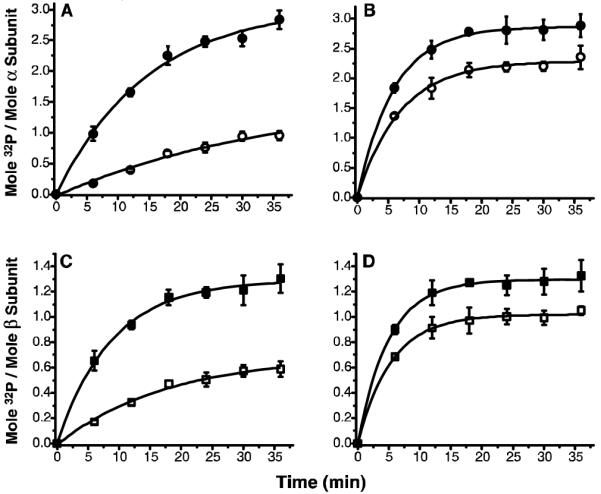
Time- and pH-dependent autophosphorylation of PhK ± acarbose. Total phosphate incorporation into the α and β subunits of PhK in the absence (open symbols) and presence (closed symbols) of acarbose (250 μM) at pH 6.8 (A&C) and pH 8.2 (B&D) was measured on P81 filters. In parallel, at indicated intervals, aliquots of the reaction mixture were run on 7.5%T SDS-PAGE. The 32P incorporation into each subunit was quantified on a Typhoon 9410 Phosphor Imager (Amersham Biosciences, Piscataway, NJ) following autoradiography of the gels. An asymptotic function y=a−bcx was applied for exponential fitting (p<0.05) of each data set using Origin 7.5 software (OriginLab, Northampton, MA).