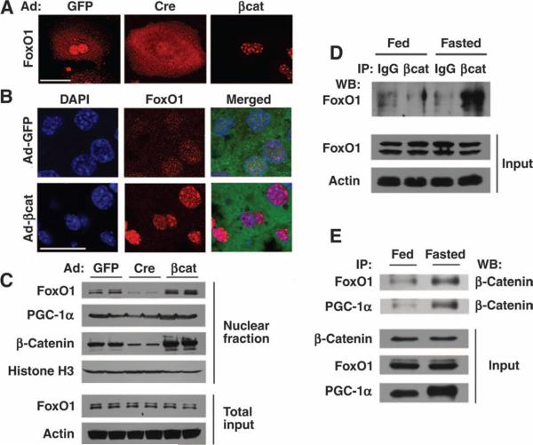
Fig. 2.
Interaction of FoxO1 with β-catenin. (A) Primary hepatocytes isolated from β-catenin floxed mice were infected with the indicated adenoviruses, and FoxO1 subcellular localization was subsequently assessed by indirect immunofluorescence. (B) In vivo localization of FoxO1 was determined in mice previously infected with the indicated adenoviruses and starved overnight. The Ad-βcat vector is a bicistronic construct that also encodes GFP. (C) Western blot analysis for the abundance of nuclear FoxO1, PGC-1α, and β-catenin. Nuclear fractions were prepared from mice previously infected with the indicated adenoviruses. Histone H3 was used as a loading control for nuclear proteins and actin as a control for the total hepatic lysate. (D) Lysates were prepared from whole livers of either fed mice or animals starved for 18 hours. Equal amounts of protein were immunoprecipitated (IP) with an antibody directed against β-catenin or with a control IgG serum, and the amount of coprecipitating FoxO1 was assessed by Western blot (WB) analysis. (E) Nutrient-sensitive protein interactions in fed and fasted livers observed between β-catenin and both FoxO1 and PGC-1a. Scale bar in immunohistochemical panels, 20 μm. All Western blots are representative of experiments that were performed at least three times.

