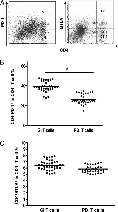
Fig. 2.
PD-1 and BTLA expression in gastric infiltrating/peripheral blood CD4+ T cells in patients with H. pylori infection. (A) Surface expression of PD-1 and BTLA on gastric infiltrating CD4+ T cells in one patient with H. pylori infection. (B and C) Percentages of CD4+ PD-1+ and CD4+ BTLA+ gastric infiltrating (GI)/peripheral blood (PB) T cells in patients with H. pylori infection. Horizontal bars indicate the median percentages of CD4+ PD-1+ T cells and CD4+ BTLA+ T cells. The individual frequency for each subject included in the analysis is shown. Significance was calculated using the Mann-Whitney U test. *, P < 0.05.

