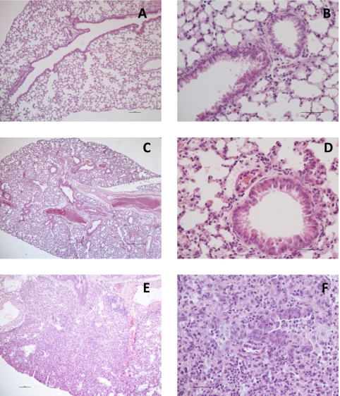
Fig. 3.
Histopathology on different mouse strains challenged with H1N1 PR8 virus with or without pretreatment with IgY to H5N1. (A and B) C.B-17 uninfected control; normal lungs. (C and D) C.B-17 mouse treated with IgY against H5N1 and challenged with 104 TCID50 PR8. (A and C) The lungs do not show any inflammatory lesions. (B and D) Intact bronchiolar mucosa and alveoli are visible. (E and F) C.B-17 mouse treated with control IgY and challenged with 104 TCID50 PR8. (E) Severe pulmonary inflammation. (F) The bronchiolar mucosa is damaged and infiltrated with leukocytes, predominantly polymorphs and macrophages. Leukocytes in the lumen and the peribronchial tissues are also visible. Magnifications, ×60 (A), ×240 (B), ×24 (C), ×240 (D), ×60 (E), and ×240 (F).

