Fig. 2.
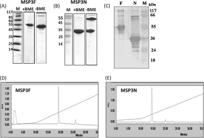
(A and B) Coomassie blue-stained SDS-polyacrylamide gel showing purified MSP3F and MSP3N under reducing and nonreducing conditions. Molecular mass markers (in kDa) are shown. BME, β-mercaptoethanol. (C) Western blot by human hyperimmune sera shows recognition of both recombinant proteins MSP3F (lane F) and MSP3N (lane N). Lanes M, molecular size markers. (D and E) RP-HPLC profiles of purified MSP3F (D) and MSP3N (E). AU, absorbance units.

