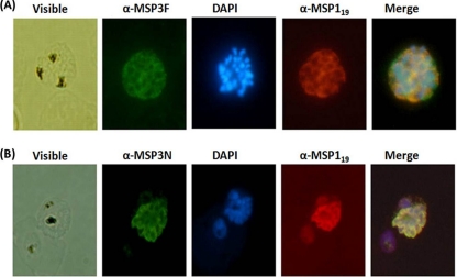
Fig. 5.
Immunofluorescence assay with P. falciparum 3D7 parasites using anti-MSP3F (A) (green) and anti-MSP3N (B) (green). Both panels also include immunostaining with anti-MSP119 antibodies (red). Shown are fluorescence and bright-field images of acetone-methanol-fixed P. falciparum 3D7 parasites at the schizont stage. Parasite nuclei were stained with DAPI (blue).

