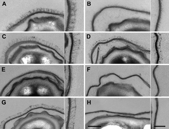
Fig. 1.
Ultrathin sections of B. cereus ATCC 14579 and B. anthracis 9131 spores and the related mutants visualized by transmitted electron microscopy (scale bar = 100 nm). For each strain, the inset is an enlargement of a representative part of the corresponding exosporium (scale bar = 50 nm). (A to F) B. cereus ATCC 14579. (A) Wild type. (B) ΔbclA. (C) ΔbclA pHT304 bclA. (D) ΔexsH. (E) ΔbclA ΔexsH. (F) ΔbclA ΔexsH pHT304 exsH. (G and H) B. anthracis 9131. (G) Wild type. (H) ΔbclA.

