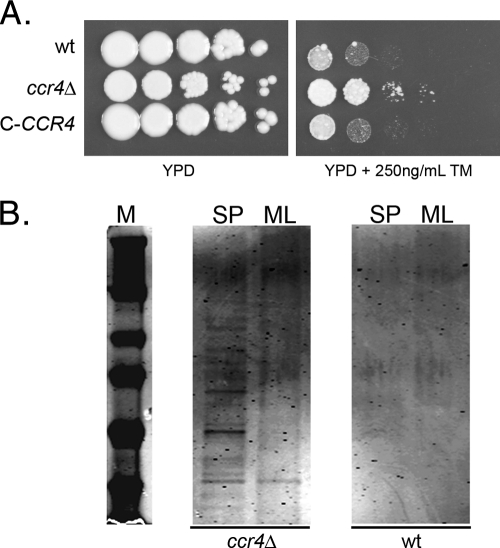
Fig. 1.
(A) Spot plate assay comparing the tunicamycin (TM) sensitivities of the wild type, the ccr4Δ mutant, and a complemented mutant (C-CCR4). The spots are 5 μl of a suspension at an OD600 of 1.0 and 4 serial 10-fold dilutions. The plates were incubated at 30°C for 3 days and photographed. (B) SDS-PAGE analysis of supernatants from 20 OD600 units of cells from mid-log-phase (ML) or stationary-phase (SP) cultures of the wild type and the ccr4Δ mutant incubated overnight at 4°C in mild DTT extraction buffer. The gel was stained and imaged on an Odyssey IR scanner (Li-Cor).

