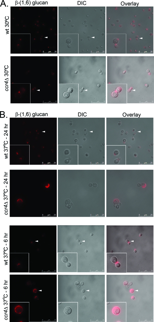
Fig. 3.
Localization of β-(1,6) glucan on the surfaces of C. neoformans wild type and the ccr4Δ mutant. Cells were grown overnight at 30°C or 37°C or shifted to 37°C for 6 h, formaldehyde fixed, and stained with a rabbit polyclonal anti-β-(1,6) glucan antiserum, followed by an Alexa Fluor 586-conjugated secondary antibody. The arrowheads indicate the regions of the images expanded in the insets. DIC, differential interference contrast.

