Fig. 4.
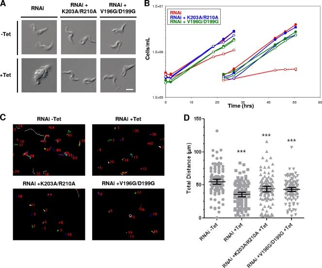
Bloodstream-form motility mutants are viable. (A) DIC images of live BSF cells grown with or without Tet for 48 h, as indicated. Tet-induced LC1 3′ UTR RNAi cells (RNAi) are inviable and accumulate as amorphous masses with multiple flagella, indicating cytokinesis failure. LC1 mutants with point mutations (RNAi + K203A/R210A and RNAi + V196D/D199G) are viable and have normal morphology. Bar, 5 μm. (B) Growth curves of bloodstream-form LC1 3′ UTR RNAi knockdowns (RNAi, red lines) or LC1 mutants with point mutations (blue lines, K203A/R210A; green lines, V196D/D199G), demonstrating that LC1 mutants with point mutations rescue the lethal phenotype caused by LC1 knockdown. Cultures were incubated with (open symbols) or without (closed symbols) tetracycline added at time zero, and cells were diluted back to the starting density at 25 h. (C) Motility traces of control cells (RNAi − Tet), LC1 UTR knockdowns (RNAi + Tet), and LC1 mutants with point mutations (RNAi + K203A/R210A and RNAi + V196G/D199G) were carried out as described in the legend to Fig. 2C (the motility of mutants with point mutations in the absence of Tet was like that of the wild type and is not shown). (D) Quantification of total distance traveled by individual cells in motility traces from panel C (N > 100 for each culture) was carried out as described in the legend to Fig. 2D. Horizontal lines indicate the mean of each data set, with bars indicating the 95% confidence interval. Results for the data sets were compared to those for RNAi without Tet. ***, significant difference (P < 0.0002).

