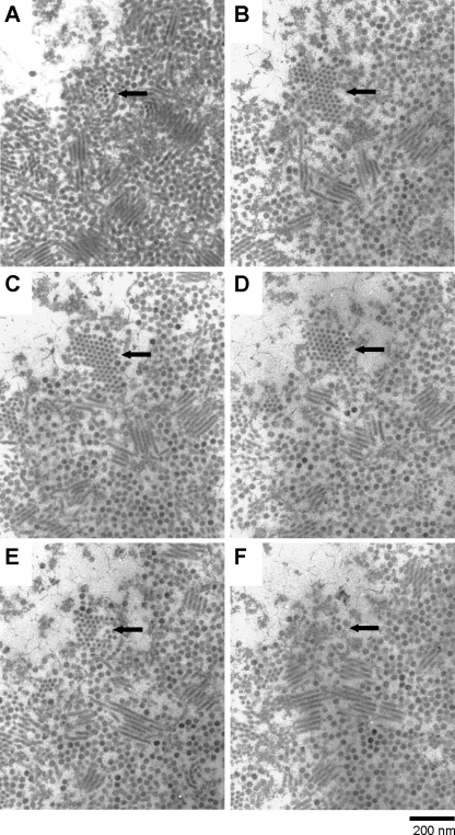
Fig. 4.
Transmission electron micrographs of continuous serial gray-colored ultrathin sections (A to F) of a virus-like particle and an electron-dense rod-shaped particle assemblage site in the nucleus of a ClorDNAV-infected Chaetoceros lorenzianus cell (48 hpi). The predicted thickness of each section is <60 nm. The top of the rod-shaped particle assemblage is shown in panel A, and the end is shown in panel F. Arrows indicate the same position of a horizontal section for a gathering of rod-shaped particle in the infected cell nucleus.

