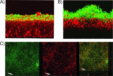
Fig. 1.
Biofilms formed in closed (A and B) and open (C) horizontal flow cells, showing donors (red), recipients (green), and transconjugants (yellow). (A) Side view of a submerged 68.5-h-old recipient MG1655::gfp biofilm observed 2.5 h after donor MG1655(pB10::rfp) was inoculated (1 h of static conditions and 1.5 h with medium flow). (B) Side view of a submerged 75.5-h-old donor MG1655(pB10::rfp) biofilm observed 4.5 h after recipient MG1655::gfp was inoculated (1 h of static conditions and 3.5 h of medium flow). Similar results were obtained when the donor and recipient cells were inoculated together and incubated for 90 h (data not shown). (C) Air-liquid interface biofilm formed 90 h after inoculation of the same donor and recipient (applied together) and transferred onto a microscope coverslip. (Left) GFP channel; (center) RFP channel; (right) both channels.

