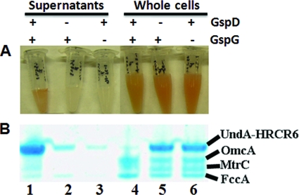
Fig. 3.
Influence of the deletion of MR-1 gspD and gspG on the extracellular release of nonacylated UndAHRCR-6. (A) Concentrated supernatants (lanes 1 to 3) and whole cells (lanes 4 to 6) of wild-type MR-1 (lanes 1 and 4), the gspD mutant (lanes 2 and 5), and the gspG mutant (lanes 3 and 6) in which nonacylated UndAHRCR-6 was overexpressed. (B) Heme staining. A total of 1 μg of concentrated supernatant proteins (lanes 1 to 3) and 10 μg of whole-cell lysate proteins (lanes 4 to 6) from the MR-1 wild type (lanes 1 and 4), gspD mutant (lanes 2 and 5), and gspG mutant (lanes 3 and 6) were separated by SDS-PAGE and then visualized by heme staining.

