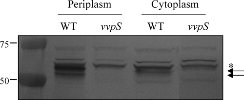
Fig. 6.
VvpS in the cytoplasmic and periplasmic spaces. Cytoplasm and periplasm fractions of the wild type and vvpS mutant grown in LBS for 24 h were prepared by using the PeriPreps Periplasting Kit (Epicentre Biotechnologies, Madison, WI). The resulting fractions, equivalent to 10 μg of total proteins, were loaded in each lane and examined for the presence of VvpS protein by Western blot analyses. The Western blots are presented as described in the legend to Fig. 2. Molecular mass markers (Precision Plus Protein standards; Bio-Rad Laboratories) for VvpS proteins (arrows) and a cross-reacting protein (asterisk) are shown in kilodaltons.

