Fig. 1.
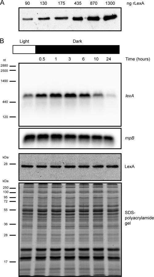
LexA is regulated on a posttranscriptional level. (A) The dynamic range of the anti-LexA antibodies raised in the present work was tested using purified rLexA; the amount of rLexA loaded on each well is shown. (B) Synechocystis sp. strain PCC 6803 cells were grown in BG11 under continuous light before being transferred to dark conditions. Cells were collected at different time points, and total RNA and proteins were extracted from the samples. Northern blot analyses of the relative levels of lexA and rnpB transcripts are shown (two upper blots). The numbers on the left of the upper blot indicate sizes in nucleotides as estimated from the rRNA and rnpB bands. The two lower panels depict the Western blot analysis of LexA and the Coomassie blue-stained SDS-polyacrylamide gel showing the equal loading of the samples. The molecular masses of the Fermentas protein marker are indicated on the left.

