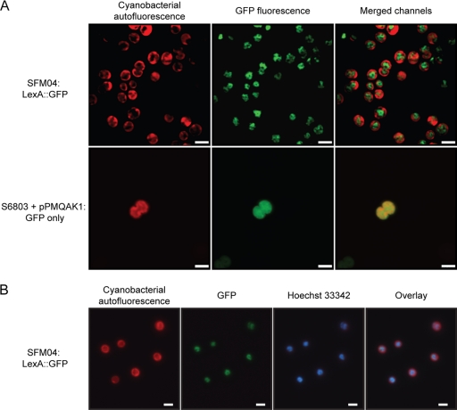
Fig. 7.
LexA::GFP in SFM04 is located in the core region of the cytoplasm in an evenly distributed pattern. (A) Confocal micrographs were obtained from cells of the strain SFM04 (possessing the LexA::GFP fusion protein) (top row) and of Synechocystis sp. strain PCC 6803 harboring the self-replicative plasmid pPMQAK1, which contains gfp expressed under the regulation of a modified trc promoter (19) (bottom row). The cyanobacterial autofluorescence is depicted in the left column, while the collected GFP signal is shown in the middle column. The result of merging the signals from both channels (cyanobacterial autofluorescence and GFP) is shown in the right column. Size bars, 2.5 μm. (B) Fluorescence micrographs were acquired from SFM04 cells, specifically analyzing the cyanobacterial autofluorescence, the LexA::GFP signal, and the fluorescence of the DNA-staining dye Hoechst 33342. An overlay of the different micrographs is shown on the right. Size bars, 2 μm.

