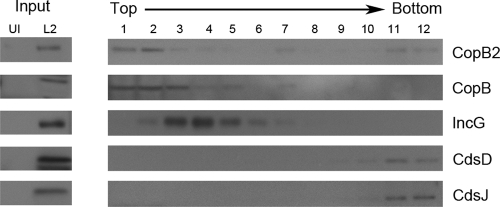
Fig. 3.
CopB and CopB2 fractionation in whole-culture membrane flotation gradients. C. trachomatis-infected (L2) or mock-infected (UI) HeLa cultures were disrupted 24 h postinfection, and membrane-containing, clarified lysates (input) were generated. Lysates were subjected to density gradient centrifugation through sucrose. Twelve individual column fractions were collected, and proteins were concentrated via TCA precipitation. Equivalent volumes of material corresponding to the top (1) to the bottom (12) fractions of the column were resolved by SDS-PAGE in 12% polyacrylamide gels. Immunoblots of input and column fractions were probed with anti-CopB and anti-CopB2 or with anti-IncG, anti-CdsD, and anti-CdsJ as fractionation controls. Proteins were visualized by probing with secondary antibodies conjugated to horseradish peroxidase and development with ECL Plus chemiluminescence reagent.

