Fig. 5.
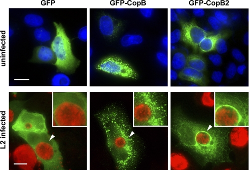
GFP-CopB2 colocalizes with inclusions when ectopically expressed in C. trachomatis-infected HeLa cells. pGFP-CopB and pGFP-CopB2 as well as the negative control, pEGFP, were transfected into uninfected or C. trachomatis L2-infected HeLa cells. Cultures were fixed at 18 h posttransfection, and localization of recombinant proteins (EGFP, GFP-CopB, and GFP-CopB2; green) was observed. HeLa cell nuclei of uninfected cultures were visualized by staining with DAPI (blue), whereas chlamydiae (red) in infected cultures were visualized with anti-GroEL and Alexa-594-conjugated secondary antibodies. Images were acquired via epifluorescence microscopy, and arrows indicate areas presented in insets. Scale bar, 10 μm.

