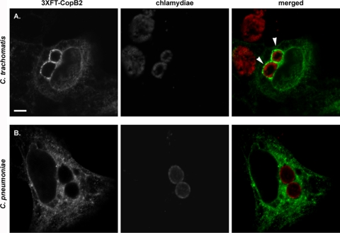
Fig. 6.
3XFT-CopB2 colocalization with C. trachomatis, but not C. pneumoniae, inclusions. HeLa cells were transfected with p3XFT-CopB2 after infection with C. trachomatis L2 (A) or C. pneumoniae AR-39 (B) and processed for microscopy at 18 h posttransfection. These times corresponded to ca. 24 h postinfection for C. trachomatis and 64 h for C. pneumoniae. Recombinant protein was detected using anti-FLAG (green), and chlamydiae were detected using MOMP-specific antibodies (red). Proteins were visualized by probing with appropriate secondary antibodies and viewed using confocal laser scanning microscopy. Arrows designate colocalization of 3XFT-CopB2 with chlamydial inclusions. Scale bar, 5 μm.

