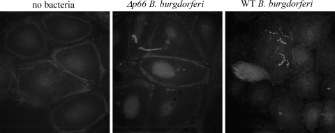
Fig. 5.
P66 disrupts cortical actin in epithelial cells. HEK 293 cells transfected to express integrin αvβ3 were infected at an MOI of 10 with p66+ or Δp66 B. burgdorferi. The infections were allowed to proceed at 37°C under 5% CO2 for 1 h, after which the cells were washed to remove unbound bacteria and fixed in paraformaldehyde. After permeabilization in 0.1% Triton X-100, the samples were stained with anti-OspA mouse serum plus Alexa Fluor 350-phalloidin. Note that in the cells infected with the bacteria that express P66, the cortical rim of actin has largely dissipated, and the cells appear to have contracted. All panels are shown at the same magnification.

