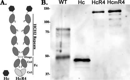
Fig. 2.
Structure and expression of recombinant Hc antigens. (A) Schematic of recombinant Hc and HcR4. HcR4 consists of Hc expressed as a fusion protein with R4, a tandem repeat composed of 4 copies of the hinge and CH2 sequences (HCH2) from IgG fused to an Fc region of IgG. The HCH2 sequence contains the region of IgG that binds to FcγRs. (B) Western blot analysis of Hc antigens. Proteins were separated on SDS-PAGE gels under reducing conditions and transferred to nitrocellulose membranes. Hc antigens were detected by using an anti-BoNT/A1 polyclonal antibody. Lane 1, wild-type BoNT/A1 Hc (WT; 200 ng); lane 2, recombinant Hc (50 ng); lane 3, HcR4 (150 ng); lane 4, HcmR4 (150 ng). Positions of molecular weight markers, in thousands, are shown to the left.

