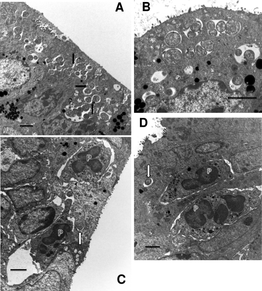
Fig. 2.
Events occurring 24 h after infection. (A) Multiple early inclusions containing one to a few RBs. Some epithelial cells contain multiple inclusions. RBs are clearly dividing, and the proximity of inclusions suggests that the fusion process is under way (arrows). (B) Enlarged view of early, 24-h inclusions clearly showing multiple inclusions in a single cell. The RBs are generally larger at this time of the cycle in preparation for division than at later times. (C) PMNs (P) are seen directly in contact with an infected cell even though only a single RB (arrow) is present. Note the obvious pseudopodia of the PMNs extending toward and actually forming pockets in the infected cell. (D) Another view showing PMNs (P) in contact with an infected cell containing a single RB (arrow). Scale bars: 2 μm.

