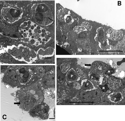
Fig. 3.
Events at 30 and 36 h after infection. (A) Inclusion with multiple RBs at 30 h after infection and PMNs (P) in contact with the infected epithelial host cell. (B) By 36 h after infection, the inclusions have increased dramatically in size and contain plentiful EBs, indicating that the inclusion is in its late stages. PMNs (P), again, abut the infected epithelia. (C) An infected cell, with PMNs (P) on either side (arrow), is becoming detached from the epithelium. Note that the inclusion membrane is no longer apparent and the chlamydiae appear to be free in the cytoplasm. The highly vacuolated nature and lack of microvilli of the infected cell suggest that the epithelial cell is dying. (D) Multiple large mature inclusions are seen. In particular, one infected cell (arrow) is being detached from the epithelium with a PMN (P) directly in the space beneath the cell, giving the appearance of “pushing” the cell off the epithelial surface; the PMN appears to have phagocytized numerous RBs. Nuclei (N) of the infected cells are visible. Scale bars: 2 μm (A and C), 10 μm (B and D).

