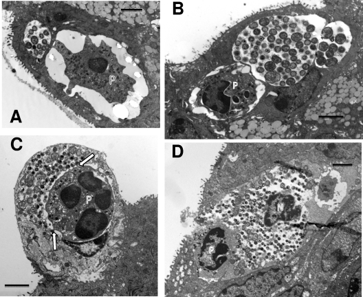
Fig. 4.
PMNs enter into the infected host epithelial cell to make direct contact with chlamydiae. (A) At 42 h after infection, a PMN has gained access to and is completely inside the infected target cell. Note the extended pseudopodia, one directly in contact with the epithelial cell cytoplasm close to the inclusion. (B) Another view showing an invading intracellular PMN directly in contact with the chlamydial inclusion membrane. (C) The inclusion membrane is no longer apparent, and the intracellular PMN is in direct contact with chlamydiae. Note the projections from the PMNs touching the IBs or EBs (arrows). (D) A PMN (P) within a large space containing chlamydiae and cellular debris, including a cell nucleus (N), suggesting either that this epithelial cell was destroyed by the action of the PMN or that the PMN has entered the site following lysis of the cell. Scale bars: 2 μm (A and C), 10 μm (B and D).

