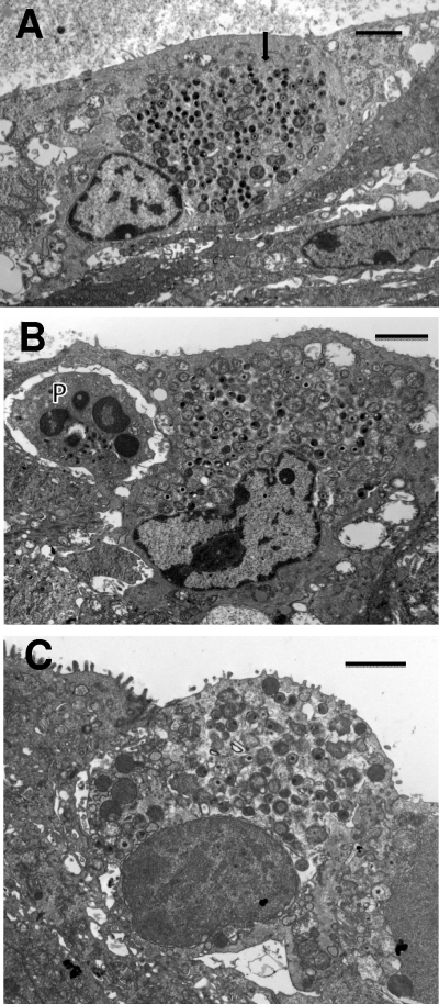
Fig. 5.
Termination of the developmental cycle by breakdown of the inclusion membrane. (A) Host epithelial cell with multiple RBs and EBs distributed in the cytoplasm (arrow). The inclusion membrane is no longer obvious. (B) Similar micrographs showing a terminal infected cell with chlamydiae freely distributed in the cytoplasm. A PMN (P) is adjacent to the infected cell. (C) Terminal infected cell with chlamydiae distributed in the cytoplasm. The cell is in the process of being dislodged from the epithelial layer. Scale bars: 2 μm (A and C), 10 μm (B).

