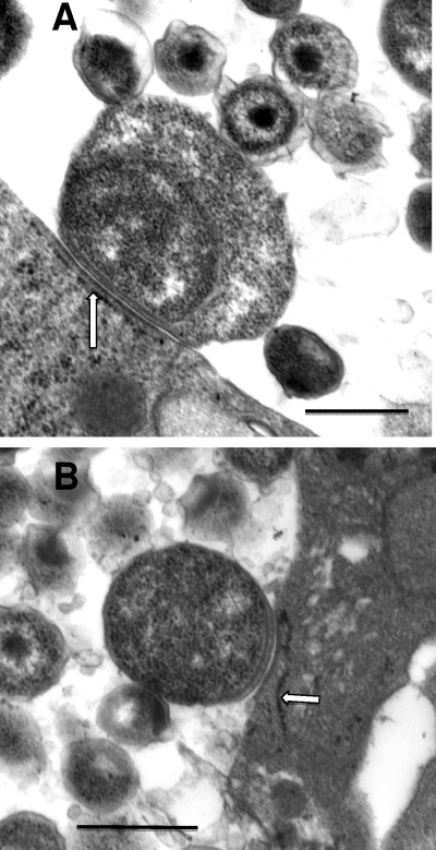
Fig. 7.
RBs attached to the inclusion membrane reflecting the contact-dependent hypothesis. (A) Enlarged replicative RB with extensive inclusion membrane contact, likely via the type 3 secretion injectisomes; the contact areas are almost always associated with closely apposed endoplasmic reticulum (arrow), from which the chlamydiae are likely scavenging nutrients. (B) Smaller RB with less extensive area of contact with the inclusion membrane. Scale bars: 500 nm.

