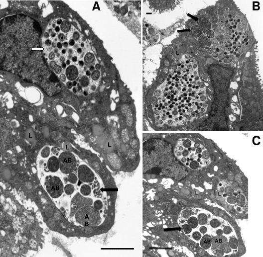
Fig. 9.
Inclusions containing aberrant RBs were observed in the cervix of a mouse depleted of PMNs by antibody treatment. (A) Note the large aberrant RBs (AB) as well as packets of miniature RBs (black arrow). Also, this inclusion is devoid of granular material representing glycogen accumulation (white arrow). (B) A large mature inclusion is in the lower left of the photomicrograph, while the upper right inclusion has both aberrant RBs (arrows) and normal size RBs as well as IBs and EBs. (C) Another view showing a normal inclusion (top center) and two inclusions with aberrant RBs (AB), packets of miniature reticulate bodies (arrow), and a lack of glycogen. Scale bars: 2 μm (A and C), 500 nm (B).

