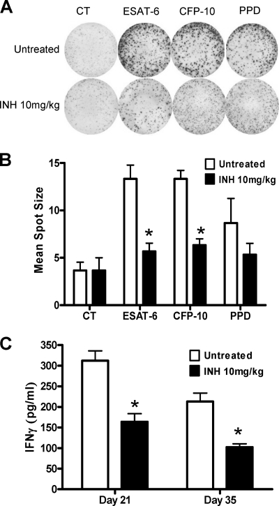
Fig. 4.
Impact of isoniazid treatment on IFN-γ response in vitro and in vivo. (A) The densities of spot-forming cells before and after isoniazid treatment at day 35 are shown for ESAT-6, CFP-10, and PPD stimulation in duplicate at 10 mg/ml. (B) The mean spot sizes (in square millimeters) in the IFN-γ ELISpot assay are compared between stimulated PBMCs from the isoniazid-treated (black bars) and untreated (white bars) infected animals. The mean spot sizes (from duplicates of each animal) and SDs are shown. (C) Serum IFN-γ measured by ELISA 2 weeks and 4 weeks after isoniazid treatment start. Four animals were used at each time point, and the mean spot sizes or serum IFN-γ concentrations are shown. *, P < 0.05 (Student's t test), comparison between treated and untreated groups. The experiment was carried out twice independently. CT, control (unstimulated) group.

