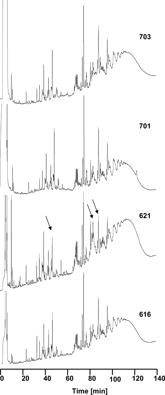
Fig. 2.

Muropeptide pattern. The cell wall of all four strains was isolated and digested by muraminidase mutanolysin and analyzed by HPLC. The muropeptide patterns of strains 616, 701 and 703 were always very similar. Differences in the muropeptide pattern of strain 621 compared to those of the other three strains are indicated by black arrows.
