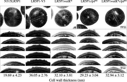
Fig. 3.
Transmission electron microscopy of the gene replacement mutants and their control strains. Transmission electron microscopy was carried out on gene replacement mutants LR5P1walK* and LR5P1clpP*, which were generated from N315LR5P1 by substitution of its walK and clpP genes with those of LR5P1-V3, respectively. The LR5P1walK*clpP* mutant was generated from LR5P1walK* by substitution of its clpP gene with that of LR5P1-V3. The values given under each picture are the means and standard deviations of each strain's cell wall thickness. Note that all gene replacement cells had thick cell walls compared to those of the parent strain. Magnifications, ×30,000.

