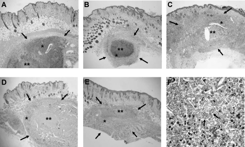
Fig. 3.
Histological sections of the nodular lesions in athymic mice 4 months after challenge with 3 × 10⁶ CFU of F. pedrosoi FMR 6630. Control mice (A) and mice treated with VRC at 20 mg/kg (B) show subcutaneous nodules (delimited by arrows) with important inflammatory responses (*) and central zones of necrosis (**). Mice treated with TRB at 250 mg/kg (C), mice treated with ITZ at 50 mg/kg (D), and mice treated with PSC at 20 mg/kg (E) show smaller subcutaneous nodules with central necrosis but with a less intense inflammatory response (H&E staining; magnification, ×25). (F) Histological nodule section showing fungal elements with sclerotic bodies (PAS stain; magnification, ×200).

