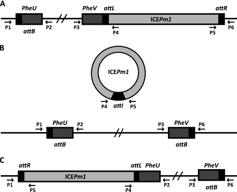
Fig. 1.
Locations of primers used in this study to detect ICEPm1 integration and excision. Oligonucleotide sequences are listed in Table 2. pheU and pheV are located on opposite DNA strands, share an identical nucleotide sequence, and are 73 bp in length. Boxes represent the 52-bp direct repeat (black), phenylalanine tRNA genes (dark gray), and ICEPm1 (light gray). The 3 possible conformations of ICEPm1 are as follows: integrated into pheV (A), excised from the chromosome and not integrated into either phe tRNA (B), and integrated into pheU (C). The figure is not to scale.

