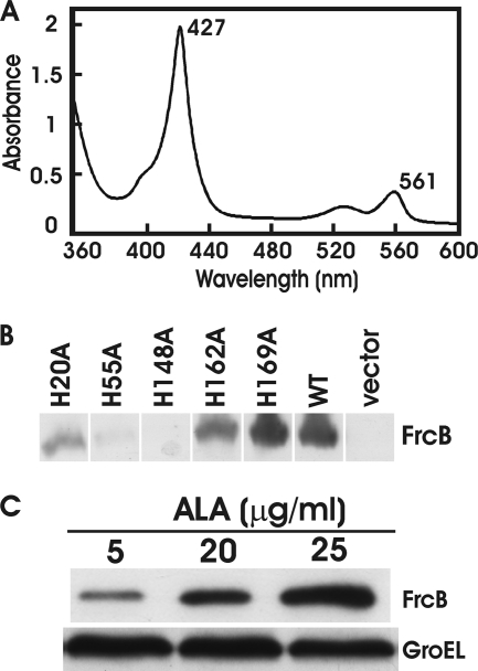
Fig. 3.
frcB encodes a diheme protein, and heme-bound FrcB was analyzed. (A) The absorption spectrum of purified, recombinant FrcB reduced with sodium dithionite by light between wavelengths of 360 and 600 nm. Peaks at 427 and 561 nm are noted. (B) Analysis of FrcB and histidine-to-alanine (His-to-Ala) mutants in E. coli cells by immunoblot analysis. An HRP-conjugated anti-His probe was used to detect His-tagged proteins. (C) Analysis of FrcB accumulation in E. coli hemA strain S905 whole cells, grown in media supplemented with 5, 20, or 25 μg/ml δ-aminolevulinic acid (ALA), by immunoblot analysis. An HRP-conjugated anti-His probe was used to detect His-tagged proteins. GroEL was used as a control for a protein not controlled by heme.

